Murray Hill Orthodontics
Contact
Hours
<ul id=”hours” style=”transition: height .3s ease;”>
<li>Monday: 9:00am – 9:00pm</li>
<li>Tuesday: 9:00am – 6:00pm</li>
<li>Wednesday: 9:00am – 9:00pm</li>
<li>Thursday: 9:00am – 9:00pm</li>
<li>Friday: 9:00am – 5:00pm</li>
</ul>

Murray Hill Orthodontics, located in the heart of New York, New York, is dedicated to providing exceptional orthodontic care in a warm and welcoming environment. Led by a team of experienced orthodontists, our practice combines personalized treatment plans with state-of-the-art technology to help patients achieve beautiful, healthy smiles. Whether you’re interested in traditional braces or clear aligners, Murray Hill Orthodontics offers a comprehensive range of orthodontic services to suit your needs.
Orthodontic Services
Traditional Braces
- Metal Braces: Time-tested orthodontic solution for correcting misaligned teeth and bite issues.
- Ceramic Braces: Clear or tooth-colored brackets for a more discreet orthodontic treatment option.
Clear Aligner Therapy
- Invisalign: Removable clear aligners for a virtually invisible way to straighten teeth without traditional braces.
- ClearCorrect: Transparent aligners that gradually shift teeth into alignment for a more aesthetically pleasing smile.
Comprehensive Orthodontic Care
Initial Consultation
- Orthodontic Evaluation: Thorough assessment to determine the most suitable treatment plan for your individual needs and goals.
- Customized Treatment Plan: Tailored orthodontic approach designed to achieve optimal results while considering your lifestyle and preferences.
Orthodontic Treatment
- Braces Adjustment: Regular visits to ensure braces or aligners are properly adjusted for consistent progress and comfort.
- Interceptive Orthodontics: Early intervention to address orthodontic issues in children and prevent more serious problems from developing.
Patient Experience
Comfortable Environment
- Modern Facility: State-of-the-art office equipped with advanced orthodontic technology for efficient and effective treatment.
- Relaxing Atmosphere: Comfortable and inviting environment to help patients feel at ease during their orthodontic appointments.
Personalized Care
- Individualized Attention: Dedicated orthodontic team committed to providing personalized care and addressing your specific concerns throughout your treatment journey.
- Open Communication: Transparent communication about your treatment plan, progress, and any adjustments needed to ensure the best possible outcome.
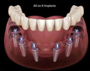
All-on-6 Dental Implants
"All-on-6" dental implants are a type of full-arch dental restoration that is supported by six dental implants strategically placed in the jawbone to support a fixed prosthesis. This treatment concept is similar to the "All-on-4" and "All-on-8" dental implant procedures but involves the placement of six implants per arch (either upper or lower) to provide stability and support for the restoration.
Here's an overview of the All-on-6 dental implant procedure:
- Evaluation and Treatment Planning:
- Before undergoing All-on-6 dental implant treatment, the patient will undergo a comprehensive dental examination, including clinical assessment and radiographic evaluation (such as dental X-rays or CBCT scans).
- The dentist or oral surgeon will assess the patient's oral health, bone density, and suitability for implant placement.
- A detailed treatment plan will be developed based on the patient's individual needs and goals.
- Implant Placement:
- The first step in the All-on-6 dental implant procedure involves surgically placing six dental implants into the jawbone at strategic locations to support the full-arch restoration.
- The number and position of the implants may vary depending on factors such as bone quality, anatomy, and the specific requirements of the case.
- Implant placement is typically performed under local anesthesia or sedation to ensure the patient's comfort during the procedure.
- Healing and Osseointegration:
- After implant placement, a healing period of several months is usually required to allow for osseointegration to occur.
- During this time, the implants fuse with the surrounding bone tissue, becoming firmly anchored in the jawbone.
- Temporary restorations may be placed during the healing period to maintain aesthetics and function.
- Restoration:
- Once osseointegration is complete, the dental implants are ready to support the full-arch restoration.
- A custom-made fixed prosthesis, typically made of high-quality dental materials such as porcelain or acrylic, is fabricated to fit securely over the implants and restore function and aesthetics.
- The prosthesis is attached to the implants using special abutments or connectors, providing stability and support for chewing and speaking.
- Postoperative Care and Maintenance:
- After the All-on-6 dental implant restoration is placed, the patient will receive instructions on postoperative care and maintenance.
- Regular dental check-ups and professional cleanings are essential to monitor the health of the implants and surrounding tissues.
- Good oral hygiene practices, including brushing, flossing, and using antimicrobial mouthwash, are important for long-term success.
All-on-6 dental implants offer several advantages over traditional removable dentures, including improved stability, function, and aesthetics. With proper care and maintenance, an All-on-6 dental implant restoration can provide long-lasting and natural-looking results, enhancing both oral health and quality of life. If you are considering All-on-6 dental implant treatment, it's important to consult with a qualified dental professional to determine the best treatment plan for your individual needs.
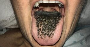
Black Hairy Tongue
Black hairy tongue is a benign and usually temporary condition characterized by a dark discoloration or "hairy" appearance of the tongue's surface. Despite its alarming appearance, black hairy tongue is typically not associated with serious health risks and can often be managed with simple interventions.
Here are some key points about black hairy tongue:
- Appearance: Black hairy tongue typically presents as elongated, dark-colored projections or "hairs" on the surface of the tongue. These projections may be black, brown, yellow, or green in color and can give the tongue a furry or hairy appearance. The discoloration is caused by the accumulation of dead skin cells, bacteria, and food debris on the papillae (tiny projections) of the tongue's surface.
- Causes: Black hairy tongue is often associated with factors that disrupt the normal shedding of dead skin cells from the surface of the tongue and allow bacteria or yeast to proliferate. Common contributing factors include poor oral hygiene, smoking or tobacco use, certain medications (such as antibiotics, antipsychotics, or antihistamines), excessive alcohol consumption, or mouth breathing.
- Symptoms: In addition to the characteristic dark discoloration of the tongue, individuals with black hairy tongue may experience a metallic taste in the mouth, bad breath (halitosis), or a sensation of dryness or tickling on the tongue's surface. In most cases, black hairy tongue does not cause pain or discomfort, although some individuals may report mild irritation or sensitivity.
- Diagnosis: Diagnosis of black hairy tongue is typically based on the characteristic appearance of the tongue and a review of the individual's medical history and lifestyle factors. In some cases, a healthcare provider may perform a physical examination of the mouth and tongue or order additional tests to rule out other potential causes of tongue discoloration or abnormal growths.
- Treatment: Treatment of black hairy tongue usually involves simple measures to improve oral hygiene and promote the shedding of dead skin cells from the tongue's surface. This may include gently brushing the tongue with a soft toothbrush or tongue scraper, using an antimicrobial mouthwash or oral rinse, and maintaining adequate hydration. Avoiding known triggers, such as tobacco use or excessive alcohol consumption, can also help prevent recurrence of black hairy tongue.
- Prognosis: Black hairy tongue is typically a self-limiting condition that resolves with appropriate oral care and lifestyle modifications. With proper treatment, the discoloration and abnormal appearance of the tongue usually improve within a few weeks. However, some individuals may experience recurrent episodes of black hairy tongue, particularly if predisposing factors are not addressed.
In summary, black hairy tongue is a benign and usually temporary condition characterized by a dark discoloration or "hairy" appearance of the tongue's surface. While it can be alarming in appearance, black hairy tongue is typically not associated with serious health risks and can often be managed effectively with simple interventions to improve oral hygiene and lifestyle habits.