
Immediate Dental Service
Contact
Hours
- Monday: 9:00am – 9:00pm
- Tuesday: 9:00am – 6:00pm
- Wednesday: 9:00am – 9:00pm
- Thursday: 9:00am – 9:00pm
- Friday: 9:00am – 5:00pm

Immediate Dental Service, located in the heart of New York, New York, is your go-to destination for prompt and efficient dental care when you need it most. With a commitment to providing immediate relief and solutions for dental emergencies, the practice offers a comprehensive range of urgent dental services in a convenient and accessible location. Led by a team of experienced emergency dentists, Immediate Dental Service strives to deliver high-quality care with minimal wait times, ensuring that patients receive the attention they need when experiencing dental pain or discomfort.
Urgent Dental Services
Emergency Examinations
- Rapid Assessments: Immediate evaluations to diagnose dental emergencies and determine the appropriate course of action.
- Pain Management: Quick relief from dental pain through various techniques, including local anesthesia and sedation options.
Emergency Dental Treatments
- Emergency Extractions: Prompt removal of severely damaged or infected teeth causing acute pain or discomfort.
- Emergency Root Canal Therapy: Expedited root canal treatment to alleviate pain and save infected teeth.
Immediate Care
Same-Day Appointments
- Flexible Scheduling: Accommodating same-day appointments for patients experiencing dental emergencies.
- Walk-In Services: Convenient walk-in availability for urgent dental care without the need for a prior appointment.
Efficient Treatment
- Rapid Response: Prompt attention from experienced emergency dentists to address urgent dental needs without delay.
- Streamlined Procedures: Expedited processes to minimize waiting times and efficiently manage dental emergencies.
Patient Care
Compassionate Approach
- Empathetic Staff: Caring and supportive dental professionals dedicated to providing comfort and reassurance during stressful situations.
- Clear Communication: Transparent explanations of treatment options and expectations to empower patients to make informed decisions about their dental care.
Aftercare Support
- Post-Treatment Guidance: Clear instructions and guidance on managing post-procedure discomfort and maintaining oral health following emergency dental treatments.
- Follow-Up Care: Scheduled follow-up appointments to ensure the successful resolution of dental emergencies and address any lingering concerns.

Toothache Treatment
Toothaches can range from mild discomfort to severe pain and can be caused by various factors, including dental decay, gum disease, dental trauma, infection, or tooth sensitivity. While proper diagnosis and treatment by a dentist are essential for addressing the underlying cause of a toothache, there are several home remedies and over-the-counter treatments that can help alleviate pain and discomfort temporarily. Here are some toothache treatments and home remedies:
- Over-the-counter Pain Medication:
- Nonsteroidal anti-inflammatory drugs (NSAIDs) such as ibuprofen (Advil, Motrin) or naproxen (Aleve) can help reduce pain and inflammation associated with toothaches. Follow the recommended dosage instructions on the label or consult with a healthcare professional for guidance.
- Topical Analgesics:
- Over-the-counter topical analgesic gels or creams containing benzocaine or lidocaine can provide temporary relief from toothache pain by numbing the affected area. Apply a small amount of the gel directly to the sore tooth and surrounding gums as directed on the product label.
- Cold Compress:
- Applying a cold compress or ice pack to the outside of the cheek near the painful tooth can help numb the area and reduce swelling and inflammation, providing temporary relief from toothache pain. Wrap the ice pack in a cloth or towel to prevent direct contact with the skin and apply it to the affected area for 10-15 minutes at a time.
- Saltwater Rinse:
- Rinsing your mouth with warm saltwater can help reduce inflammation and kill bacteria in the mouth, providing temporary relief from toothache pain. Dissolve a teaspoon of salt in a glass of warm water and swish the solution around in your mouth for 30-60 seconds before spitting it out.
- Clove Oil:
- Clove oil contains eugenol, a natural compound with analgesic and numbing properties that can help relieve toothache pain. Apply a small amount of clove oil to a cotton ball or swab and gently dab it onto the sore tooth and surrounding gums. Be cautious not to swallow the oil or apply it directly to the tongue or other sensitive tissues, as it can cause irritation.
- Peppermint Tea Bags:
- Peppermint tea bags contain menthol, which has cooling and numbing properties that can help alleviate toothache pain. Steep a peppermint tea bag in hot water for a few minutes, allow it to cool slightly, and then place it against the affected tooth for 10-15 minutes.
- Avoid Trigger Foods and Beverages:
- Avoid consuming hot, cold, sweet, or acidic foods and beverages that can exacerbate tooth sensitivity and pain. Stick to soft, bland foods and room-temperature or lukewarm liquids until the toothache resolves.
- Maintain Good Oral Hygiene:
- Continue to practice good oral hygiene habits, including brushing with a soft-bristled toothbrush and fluoride toothpaste, flossing daily, and using an antiseptic mouthwash to help reduce bacteria and plaque buildup in the mouth.
- Avoid Applying Aspirin Directly to the Tooth:
- While some people may suggest placing a crushed aspirin tablet directly on the sore tooth to alleviate pain, this practice can cause chemical burns to the gums and other oral tissues and should be avoided.
It's important to note that these home remedies and over-the-counter treatments are intended to provide temporary relief from toothache pain and discomfort while awaiting professional dental care. If you experience severe or persistent toothache pain, swelling, fever, or other signs of infection, it's essential to seek prompt evaluation and treatment by a dentist to address the underlying cause of the toothache and prevent complications. Delaying treatment for a toothache can lead to worsening pain, infection, and possible tooth loss, so don't hesitate to schedule an appointment with your dentist for proper diagnosis and treatment.
Dental Erosion
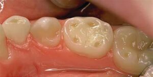
Dental erosion refers to the gradual loss of tooth enamel due to chemical processes rather than bacterial action, which is typical of dental decay. It occurs when acids, often from dietary sources or gastric reflux, come into contact with the tooth surface and dissolve the mineral content of the enamel. Dental erosion can lead to tooth sensitivity, discoloration, and an increased risk of cavities and tooth loss if left untreated.
Here are some key points about dental erosion:
- Causes:
- Acidic foods and beverages: Consuming acidic foods and drinks such as citrus fruits, carbonated beverages, fruit juices, vinegar, and sour candies can contribute to dental erosion.
- Gastric reflux: Conditions like gastroesophageal reflux disease (GERD) or bulimia, which involve frequent vomiting or regurgitation of stomach acids, can expose the teeth to acidic substances and contribute to erosion.
- Environmental factors: Exposure to environmental acids, such as industrial pollutants or chlorinated swimming pool water, can also contribute to dental erosion.
- Medications: Certain medications, such as aspirin or vitamin C supplements in chewable form, can be acidic and contribute to erosion if they come into contact with the teeth.
- Dry mouth: Reduced saliva flow, as seen in conditions like xerostomia or as a side effect of medications, can decrease the mouth's natural buffering capacity and increase susceptibility to erosion.
- Symptoms:
- Tooth sensitivity: Eroded enamel exposes the underlying dentin, which contains nerve endings, leading to increased sensitivity to hot, cold, sweet, or acidic stimuli.
- Discoloration: Teeth may appear yellowish or dull due to thinning enamel and increased visibility of the underlying dentin.
- Rounded or cupped tooth surfaces: Erosion may cause the enamel to wear away unevenly, resulting in rounded or concave areas on the tooth surface.
- Transparency: The edges of the teeth may appear translucent or see-through due to thinning of the enamel.
- Increased risk of cavities: As enamel erodes, the underlying dentin becomes more susceptible to decay, increasing the risk of cavities.
- Diagnosis:
- Diagnosis of dental erosion is typically based on clinical examination, dental history, and visual inspection of the teeth. Your dentist may also inquire about dietary habits, medical conditions, and lifestyle factors that could contribute to erosion.
- Dental X-rays or intraoral cameras may be used to assess the extent of enamel loss and evaluate the condition of the underlying dentin.
- Treatment:
- Treatment of dental erosion aims to prevent further enamel loss, alleviate symptoms, and protect the teeth from decay. Treatment options may include:
- Dietary modifications: Limiting consumption of acidic foods and beverages can help reduce acid exposure and slow the progression of erosion.
- Oral hygiene practices: Brushing with a fluoride toothpaste and using a fluoride mouth rinse can help strengthen enamel and protect against decay.
- Dental restorations: Depending on the severity of erosion and symptoms, your dentist may recommend dental bonding, veneers, or crowns to restore and protect damaged teeth.
- Saliva stimulation: Chewing sugar-free gum or using saliva substitutes can help stimulate saliva flow and improve oral pH balance to reduce acidity.
- Acid-neutralizing agents: Your dentist may recommend prescription or over-the-counter products containing calcium, phosphate, or fluoride to remineralize enamel and neutralize acids.
- Prevention:
- To prevent dental erosion, it's important to:
- Limit consumption of acidic foods and beverages, and rinse the mouth with water afterward to neutralize acids.
- Use a straw when drinking acidic beverages to minimize contact with the teeth.
- Wait at least 30 minutes to brush your teeth after consuming acidic foods or drinks to avoid further enamel damage.
- Maintain good oral hygiene habits, including regular brushing, flossing, and dental check-ups.
- Seek treatment for conditions like GERD or bulimia that contribute to acid exposure and erosion.
In summary, dental erosion is the gradual loss of tooth enamel due to exposure to acidic substances. It can lead to tooth sensitivity, discoloration, and an increased risk of cavities if left untreated. Prevention and early intervention are essential to slow the progression of erosion and protect the teeth from further damage. If you suspect dental erosion or experience symptoms such as tooth sensitivity or discoloration, it's important to consult your dentist for evaluation and appropriate treatment.