

Barnet General Medical Center
Contact
Hours
<ul id=”hours” style=”transition: height .3s ease;”>
<li>Monday: 9:00am – 9:00pm</li>
<li>Tuesday: 9:00am – 6:00pm</li>
<li>Wednesday: 9:00am – 9:00pm</li>
<li>Thursday: 9:00am – 9:00pm</li>
<li>Friday: 9:00am – 5:00pm</li>
</ul>

Barnet General Medical Center, located in Buffalo, New York, is a distinguished healthcare institution renowned for its comprehensive range of medical and dental services. The hospital is equipped with cutting-edge technology and staffed by a dedicated team of highly trained animal medical professionals, committed to delivering compassionate and innovative care to all patients.
Medical Services
General Medicine and Surgery
- Emergency Services: Open 24/7, featuring rapid response teams and the latest in emergency medical technology.
- Inpatient and Outpatient Rehabilitation: Full spectrum of rehabilitation services including physical therapy, occupational therapy, and speech therapy.
- Advanced Surgical Care: Specializing in complex surgeries including cardiovascular, neurological, and reconstructive surgery.
Specialized Departments
- Endocrinology: Comprehensive care for diabetes, thyroid disorders, and other hormonal imbalances.
- Gastroenterology: Advanced diagnostic and treatment options for digestive system disorders.
- Nephrology: Expert care for kidney diseases, including dialysis services and transplant preparation.
- Psychiatry and Mental Health: Wide range of mental health services including inpatient care, outpatient therapy, and crisis intervention.
Dental Services
Preventive Dentistry
- Comprehensive Oral Exams: Thorough examinations to detect early signs of dental issues.
- Fluoride Treatments: Preventive treatments to strengthen teeth and prevent decay.
Specialized Dental Care
- Endodontics: Expert root canal therapy to save and restore damaged teeth.
- Cosmetic Dentistry: Advanced procedures including teeth whitening, veneers, and smile makeovers.
- Oral Pathology: Diagnosis and treatment of diseases affecting the mouth, jaws, and related structures.
- Prosthodontics: Specialized care for restoring and replacing teeth with bridges, dentures, and dental implants.

Fissured Tongue
A fissured tongue, also known as lingua plicata or scrotal tongue, is a benign condition characterized by the presence of deep grooves or fissures on the dorsal (top) surface of the tongue. These grooves can vary in depth and number, giving the tongue a wrinkled or cracked appearance. The condition is usually harmless and often asymptomatic, but some individuals may experience mild discomfort or sensitivity, particularly when consuming certain foods.
The exact cause of fissured tongue is not well understood, but it is believed to have a genetic component and may be associated with certain syndromes, such as Down syndrome or Melkersson-Rosenthal syndrome. It can also occur in conjunction with other conditions like geographic tongue. Good oral hygiene practices, including regular brushing of the tongue, can help manage symptoms and prevent debris from accumulating in the fissures, which can sometimes lead to irritation or infection. If there are any concerns or persistent symptoms, it is advisable to consult a healthcare professional for further evaluation and management.
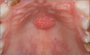
Papilloma
A papilloma is a benign epithelial tumor or growth that typically arises from the skin or mucous membranes. In the context of oral health, papillomas can occur in various locations within the oral cavity, including the lips, tongue, palate, cheeks, or gums. Oral papillomas are often caused by infection with human papillomavirus (HPV), particularly certain high-risk strains such as HPV types 6 and 11.
Here are some key points about oral papillomas:
- Types: Oral papillomas can be classified into several subtypes based on their clinical and histological features:
- Squamous papilloma: This is the most common type of oral papilloma and typically presents as a small, painless, pedunculated (stalk-like) or sessile (flat) growth on the oral mucosa. Squamous papillomas are usually pink or white in color and have a cauliflower-like appearance due to the presence of finger-like projections or papillary structures.
- Verruca vulgaris (common wart): Although more commonly found on the skin, verruca vulgaris can also occur in the oral cavity and present as papillomatous growths with a rough or hyperkeratotic surface.
- Condyloma acuminatum: This subtype of oral papilloma is associated with HPV infection and typically occurs in the anogenital region. However, it can rarely affect the oral mucosa, particularly in individuals with immunodeficiency or compromised immune function.
- Etiology: Oral papillomas are often caused by infection with HPV, a common sexually transmitted virus that can infect the skin and mucous membranes. HPV types 6 and 11 are most commonly associated with oral papillomas, particularly squamous papillomas. Transmission of HPV can occur through direct contact with infected skin or mucous membranes, including oral-genital contact or autoinoculation from other sites of infection.
- Symptoms: Oral papillomas are typically painless and may go unnoticed until they become enlarged or bothersome. Depending on their size and location, papillomas may cause discomfort, irritation, or a sensation of a foreign body in the mouth. In some cases, papillomas may bleed or become ulcerated if traumatized.
- Diagnosis: Diagnosis of oral papillomas is typically based on clinical examination and evaluation of the characteristic appearance and location of the lesion. In some cases, a biopsy may be performed to confirm the diagnosis and rule out other potential causes of oral growths.
- Treatment: Treatment of oral papillomas usually involves surgical excision or removal of the lesion, particularly if it causes symptoms, interferes with oral function, or is cosmetically undesirable. The procedure is typically performed by a dentist or oral surgeon under local anesthesia to numb the area before removal. Recurrence of oral papillomas is possible, particularly if the underlying HPV infection is not adequately treated or if predisposing factors such as immunodeficiency are present.
In summary, oral papillomas are benign epithelial tumors or growths that typically arise from the oral mucosa and are often caused by HPV infection. While usually painless and benign, papillomas can cause discomfort or interfere with oral function if they become symptomatic or enlarging. Treatment options vary depending on the size, location, and symptoms of the papilloma but may include surgical excision or removal. Early diagnosis and appropriate management are important for preventing complications and achieving favorable outcomes.

