1-888-3289

Necrotizing Sialometaplasia

At Barnet Health, we offer diagnosis and treatment of necrotizing sialometaplasia.

Necrotizing Sialometaplasia

At Barnet Health, we offer diagnosis and treatment of necrotizing sialometaplasia.

What is it?

Necrotizing sialometaplasia is an uncommon benign inflammatory condition that affects the salivary glands, particularly the minor salivary glands located in the oral cavity. It is characterized by necrosis (death) of salivary gland tissue followed by metaplasia, or the transformation of normal glandular tissue into squamous epithelium. Necrotizing sialometaplasia typically presents as a painful ulcerative lesion in the oral mucosa, often resembling a malignant neoplasm (cancer) clinically and histopathologically.

Here are some key points about necrotizing sialometaplasia:

  1. Etiology: The exact cause of necrotizing sialometaplasia is not fully understood, but it is believed to result from ischemic injury or disruption of the blood supply to the salivary glands, leading to localized tissue necrosis. Trauma, surgical procedures, local anesthesia injections, or other factors that compromise blood flow to the salivary glands may predispose individuals to develop necrotizing sialometaplasia. The condition may also be associated with systemic conditions such as vasculitis, diabetes mellitus, or autoimmune diseases.
  2. Pathogenesis: Necrotizing sialometaplasia typically begins with ischemic necrosis of the acinar cells within the salivary gland lobules, followed by reactive changes such as squamous metaplasia of the ductal epithelium. The necrotic tissue is gradually replaced by granulation tissue and fibrous scar tissue, resulting in the formation of an ulcerative lesion with raised, indurated borders. Despite its histological resemblance to malignant neoplasms such as squamous cell carcinoma, necrotizing sialometaplasia is a benign and self-limiting condition.
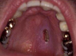
  3. Clinical Presentation: Necrotizing sialometaplasia typically presents as a painful, solitary ulcerative lesion in the oral cavity, most commonly affecting the palate, followed by the buccal mucosa (inner cheek) and floor of the mouth. The lesion may be preceded by a history of trauma or local injury and usually manifests as a well-demarcated, indurated (hardened) ulcer with irregular borders and a fibrinous or necrotic center. The surrounding mucosa may appear erythematous (red) or edematous (swollen), mimicking the clinical appearance of malignant neoplasms.
  4. Diagnosis: Diagnosis of necrotizing sialometaplasia is based on clinical examination, histopathological evaluation of a biopsy specimen, and exclusion of other potential causes of oral ulceration or malignancy. Histologically, necrotizing sialometaplasia is characterized by necrosis of salivary gland tissue, squamous metaplasia of the ductal epithelium, and the presence of granulation tissue and fibrous scar formation. Immunohistochemical staining may be performed to rule out malignant neoplasms and confirm the benign nature of the lesion.
  5. Treatment: Treatment of necrotizing sialometaplasia is typically conservative and supportive, focusing on pain management, wound care, and symptomatic relief. Nonsteroidal anti-inflammatory drugs (NSAIDs) or topical analgesics may be prescribed to alleviate pain and discomfort. Most cases of necrotizing sialometaplasia resolve spontaneously within 6-8 weeks, with complete healing and resolution of symptoms. In rare cases, surgical intervention may be necessary to debride necrotic tissue or promote wound healing in refractory or complicated cases of necrotizing sialometaplasia.

In summary, necrotizing sialometaplasia is a rare benign inflammatory condition of the salivary glands characterized by ischemic necrosis and squamous metaplasia of salivary gland tissue. Despite its clinical and histological resemblance to malignant neoplasms, necrotizing sialometaplasia is a self-limiting condition that typically resolves spontaneously with supportive care. Early diagnosis and appropriate management are important for relieving symptoms and preventing complications associated with necrotizing sialometaplasia.

Free E-Book!

Learn the Causes and Solutions to Gum Disease!