1-888-3289

Cementoblastoma

At Barnet Health, we offer diagnosis and treatment of cementoblastoma.

Cementoblastoma

At Barnet Health, we offer diagnosis and treatment of cementoblastoma.

What is it?

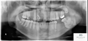
A cementoblastoma is a rare benign odontogenic tumor that arises from the cementoblasts, which are cells responsible for forming cementum, a specialized calcified tissue that covers the roots of teeth. Cementoblastomas typically occur in association with the roots of teeth, particularly the mandibular (lower) molars, and are characterized by the formation of a well-defined radiopaque (dense) mass attached to the root surface.

Here are some key points about cementoblastomas:

  1. Etiology: The exact cause of cementoblastomas is not fully understood, but they are thought to arise from aberrant proliferation or differentiation of cementoblasts within the periodontal ligament, the connective tissue that anchors teeth to the surrounding bone. Cementoblastomas are considered true neoplasms (tumors) rather than developmental anomalies, as they consist of proliferating cellular elements rather than disorganized tissue.
  2. Clinical Presentation: Cementoblastomas typically present as a painless, slow-growing swelling or mass associated with the roots of the affected tooth. The lesion is usually discovered incidentally on dental radiographs taken for unrelated reasons. On radiographic imaging, cementoblastomas appear as a well-circumscribed radiopaque mass attached to the root surface, often with a characteristic “halo” or radiolucent rim surrounding the lesion.
  3. Symptoms: In most cases, cementoblastomas are asymptomatic and do not cause pain or discomfort. However, if the lesion grows large enough, it may cause displacement or resorption of adjacent teeth, leading to localized swelling, tooth mobility, or changes in occlusion (bite). Rarely, cementoblastomas may be associated with symptoms such as toothache, facial swelling, or paresthesia (numbness or tingling) if they impinge on adjacent nerves or tissues.
  4. Diagnosis: Diagnosis of cementoblastomas is typically based on clinical and radiographic findings. Dental X-rays, including periapical, panoramic, or occlusal views, can help visualize the size, shape, and location of the lesion within the jaw. In some cases, additional imaging studies such as CT scans or MRI may be necessary to further evaluate the extent of the lesion and its relationship to surrounding structures.
  5. Treatment: Treatment of cementoblastomas usually involves surgical removal of the lesion, along with the affected tooth and surrounding periodontal tissues. The procedure is typically performed by an oral and maxillofacial surgeon and may involve extraction of the affected tooth and enucleation (surgical removal) of the tumor while preserving the surrounding bone and adjacent teeth. Following surgical removal, the prognosis for cementoblastomas is excellent, with low rates of recurrence reported.

In summary, cementoblastomas are rare benign odontogenic tumors that arise from the cementoblasts and are typically associated with the roots of teeth. While usually asymptomatic, cementoblastomas may require surgical intervention for removal if they cause symptoms or complications. Early detection and appropriate management are important for achieving favorable outcomes and preserving oral health.

Free E-Book!

Learn the Causes and Solutions to Gum Disease!