Barnet Health Doctors
Contact
Hours
- Monday: 9:00am – 9:00pm
- Tuesday: 9:00am – 6:00pm
- Wednesday: 9:00am – 9:00pm
- Thursday: 9:00am – 9:00pm
- Friday: 9:00am – 5:00pm

Barnet Health Doctors, situated in the charming town of Harris, New York, is a reputable dental institution committed to providing exceptional dental care to individuals and families in the community. With a focus on promoting oral health and delivering personalized treatment, the practice offers a wide array of dental services to address various needs and concerns. Led by a team of experienced dental professionals, Barnet Health Doctors strives to create a comfortable and welcoming environment for all patients.
Dental Services
Preventive Dentistry
- Comprehensive Examinations: Thorough dental assessments to evaluate oral health and identify any underlying issues.
- Professional Cleanings: Routine cleanings to remove plaque and tartar buildup, preventing gum disease and cavities.
- Fluoride Treatments: Application of fluoride to strengthen tooth enamel and reduce the risk of decay.
Restorative Dentistry
- Fillings: Treatment of cavities with tooth-colored composite fillings for a natural appearance.
- Crowns and Bridges: Custom-made crowns and bridges to restore damaged or missing teeth and improve functionality.
- Dental Implants: Permanent solutions for tooth replacement that mimic the look and feel of natural teeth.
Specialized Dental Care
Orthodontics
- Braces: Traditional braces and clear aligners to straighten misaligned teeth and correct bite issues.
- Retainers: Custom-fitted retainers to maintain the results of orthodontic treatment and prevent relapse.
Endodontics
- Root Canal Therapy: Treatment to remove infected or damaged pulp from within the tooth and restore its health.
- Apicoectomy: Surgical procedure to remove infected tissue from the root tip and seal the root canal.
Patient Care
Personalized Treatment Plans
- Individualized Consultations: Detailed discussions to understand each patient’s unique dental needs and goals.
- Customized Care: Tailoring treatment plans to address specific concerns and preferences.
Comfort and Convenience
- Welcoming Environment: Creating a friendly and inviting atmosphere to help patients feel at ease during their visits.
- Pain Management: Utilizing gentle techniques and effective anesthesia to minimize discomfort during procedures.
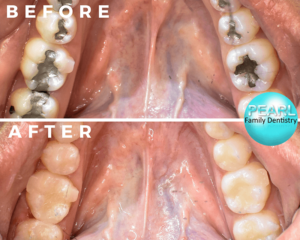
Filling Replacement
Dental filling replacement is a procedure aimed at replacing old or damaged dental fillings with new materials to restore the integrity and functionality of the affected teeth. Over time, dental fillings may wear down, become discolored, or develop cracks, which can compromise their effectiveness and lead to further dental problems if left untreated.
During a dental filling replacement procedure, the dentist will first assess the condition of the existing fillings through visual inspection and dental imaging techniques such as X-rays. If the fillings are found to be deteriorating or failing, the dentist will recommend their replacement.
The process typically involves the following steps:
- Removal of Old Fillings: The dentist will carefully remove the old fillings using dental instruments such as drills or ultrasonic scalers. This process may involve the removal of any decayed or damaged tooth structure surrounding the filling.
- Preparation of Tooth: Once the old fillings are removed, the tooth is cleaned and prepared to receive the new filling material. This may involve shaping the tooth and removing any remaining decay or debris.
- Placement of New Filling: The dentist will select an appropriate filling material based on the location and extent of the restoration needed. Common filling materials include composite resin, amalgam, porcelain, and gold. The chosen material is then placed and shaped to restore the natural contour and function of the tooth.
- Finishing and Polishing: After the new filling is placed, the dentist will carefully polish it to ensure a smooth and natural-looking surface. This helps to improve the aesthetics of the restoration and minimize the risk of plaque accumulation and staining.
- Evaluation: Once the procedure is complete, the dentist will evaluate the new filling to ensure proper fit, function, and occlusion. Any necessary adjustments may be made to achieve optimal results.
Replacing old or damaged dental fillings is essential for maintaining oral health and preventing further dental problems such as decay, infection, or fracture of the tooth. By addressing deteriorating fillings promptly, patients can preserve the strength and integrity of their teeth and enjoy long-term dental wellness. Regular dental check-ups and preventive care are key to identifying and addressing filling replacement needs in a timely manner.
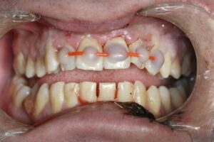
Dental Splints
Dental splints are orthodontic appliances used to stabilize, support, or realign teeth, jaws, or temporomandibular joints (TMJ). They are commonly prescribed by dentists or orthodontists to address various dental issues and provide therapeutic benefits. Dental splints come in different types and designs, each serving specific purposes and tailored to individual patient needs. Here's an overview of dental splints and their key aspects:
- Indications for Dental Splints:
- Dental splints may be recommended for patients with the following conditions:
- Teeth grinding or clenching (bruxism), which can lead to tooth wear, jaw pain, and muscle tension.
- Temporomandibular joint (TMJ) disorders, such as TMJ pain, clicking, popping, or dysfunction.
- Tooth mobility or trauma resulting from accidents, injuries, or periodontal disease.
- Orthodontic problems, including tooth misalignment, malocclusion, or bite irregularities.
- Postoperative stabilization following dental procedures, such as tooth extraction, dental implant placement, or orthognathic surgery.
- Types of Dental Splints:
- There are several types of dental splints available, each serving specific therapeutic purposes:
- Nightguards: Nightguards, also known as occlusal splints or bite guards, are custom-made appliances worn during sleep to protect the teeth and jaws from the harmful effects of bruxism. They help alleviate symptoms of teeth grinding, reduce tooth wear, and relieve jaw muscle tension.
- TMJ Splints: TMJ splints, also called occlusal appliances or bite plates, are designed to reposition the jaw and provide support for patients with TMJ disorders. They help reduce joint pain, improve jaw function, and promote relaxation of the jaw muscles by stabilizing the bite and reducing excessive force on the TMJ.
- Orthodontic Splints: Orthodontic splints, such as space maintainers, retainers, or aligners, are used to support orthodontic treatment and facilitate tooth movement. They help maintain proper tooth alignment, prevent relapse, and optimize treatment outcomes in patients undergoing orthodontic therapy.
- Periodontal Splints: Periodontal splints, also known as fixed or removable bridges, are used to stabilize mobile teeth and prevent further tooth movement in patients with periodontal disease or tooth mobility. They provide support for weakened or compromised teeth by splinting them to adjacent healthy teeth or dental implants.
- Fabrication and Customization:
- Dental splints are custom-made appliances fabricated to fit the individual patient's mouth anatomy and address specific dental concerns. The fabrication process typically involves taking impressions or digital scans of the patient's teeth and jaws, which are used to create a precise replica of the oral structures. The splint is then fabricated using biocompatible materials, such as acrylic resin or thermoplastic polymers, and adjusted to achieve proper fit, comfort, and functionality.
- Treatment Planning and Monitoring:
- Before prescribing a dental splint, the dentist or orthodontist will conduct a comprehensive evaluation of the patient's dental and medical history, perform a clinical examination, and may request additional diagnostic tests, such as X-rays or jaw movement analysis. Based on the findings, a personalized treatment plan is developed to address the patient's specific needs and goals.
- Throughout the treatment process, patients are closely monitored by their dental care provider to assess treatment progress, evaluate the effectiveness of the splint therapy, and make any necessary adjustments or modifications to ensure optimal outcomes.
- Patient Education and Maintenance:
- Patients receiving dental splints are provided with instructions on proper wear and care of the appliance, including hygiene practices, maintenance routines, and potential side effects or complications to watch for. It's important for patients to follow these guidelines and attend regular follow-up appointments to monitor their oral health and address any concerns that may arise during treatment.
- Long-Term Benefits and Outcomes:
- Dental splints can provide significant therapeutic benefits and improve the overall oral health and quality of life for patients with various dental conditions. By stabilizing the teeth, jaws, or TMJ, splints help alleviate pain, reduce tooth wear, enhance oral function, and promote long-term dental stability. With proper treatment and maintenance, patients can experience lasting relief from their symptoms and enjoy a healthier, more comfortable smile.
In summary, dental splints are valuable therapeutic appliances used in dentistry to address a wide range of dental issues, from bruxism and TMJ disorders to orthodontic problems and periodontal disease. By providing support, stabilization, and realignment of the teeth and jaws, splints help improve oral health, alleviate symptoms, and enhance the overall well-being of patients.