Emergency Dental Care USA
Contact
Hours
- Monday: 9:00am – 9:00pm
- Tuesday: 9:00am – 6:00pm
- Wednesday: 9:00am – 9:00pm
- Thursday: 9:00am – 9:00pm
- Friday: 9:00am – 5:00pm

Emergency Dental Care USA, located in the vibrant city of New York, New York, is your trusted provider of immediate dental services when unexpected dental emergencies occur. Committed to delivering prompt relief and comprehensive care for dental issues that cannot wait, our practice offers a wide range of urgent treatment options in a welcoming and comfortable setting. Led by a team of experienced emergency dentists, Emergency Dental Care USA is dedicated to restoring your oral health and alleviating your discomfort as quickly as possible.
Urgent Dental Services
Emergency Examinations
- Immediate Assessments: Thorough evaluations to diagnose and address dental emergencies promptly.
- Pain Management: Quick relief from dental pain through effective anesthesia and pain management techniques.
Emergency Dental Treatments
- Emergency Extractions: Prompt removal of severely damaged or infected teeth causing acute pain or discomfort.
- Emergency Root Canal Therapy: Expedited root canal treatment to alleviate pain and save infected teeth.
Immediate Care
Same-Day Appointments
- Flexible Scheduling: Accommodating same-day appointments for patients in need of urgent dental care.
- Walk-In Services: Convenient walk-in availability for immediate assessment and treatment of dental emergencies.
Efficient Treatment
- Rapid Response: Prompt attention from skilled emergency dentists to address urgent dental needs without delay.
- Streamlined Procedures: Expedited processes to minimize waiting times and efficiently manage dental emergencies.
Patient Care
Compassionate Approach
- Empathetic Staff: Compassionate and understanding dental professionals dedicated to providing comfort and support during stressful situations.
- Clear Communication: Transparent communication about treatment options, costs, and expectations to empower patients to make informed decisions about their dental care.
Aftercare Support
- Post-Treatment Guidance: Detailed instructions and guidance on post-procedure care and pain management to promote optimal healing and recovery.
- Follow-Up Care: Scheduled follow-up appointments to monitor progress, address any concerns, and ensure the successful resolution of dental emergencies.

Dental Cysts
Dental cysts are fluid-filled sacs or cavities that develop within the jawbone or soft tissues of the oral cavity. These cysts can arise from various sources, including developmental anomalies, inflammation, or remnants of tooth structures. Dental cysts may be asymptomatic and discovered incidentally on dental radiographs, or they may cause symptoms such as pain, swelling, or tooth displacement.
Here are some key points about dental cysts:
- Types of Dental Cysts:
- Radicular cysts: Also known as periapical cysts, radicular cysts are the most common type of dental cyst and typically develop as a result of inflammation or infection in the tooth pulp (root canal system). They occur at the apex (tip) of a non-vital tooth (a tooth with a dead or necrotic pulp) and may be associated with chronic dental caries, trauma, or failed root canal treatment.
- Dentigerous cysts: Dentigerous cysts, also called follicular cysts, form around the crown of an unerupted or impacted tooth, typically the crown of an impacted wisdom tooth or an impacted canine tooth. These cysts arise from the remnants of the tooth-forming epithelial tissue (dental follicle) and may expand and cause displacement or resorption of adjacent teeth.
- Odontogenic keratocysts: Odontogenic keratocysts are aggressive and recurrent cystic lesions that originate from the remnants of the dental lamina or enamel organ. They commonly occur in the posterior mandible and may cause jaw expansion, displacement of teeth, and cortical perforation.
- Residual cysts: Residual cysts are radicular cysts that persist after the extraction of the associated tooth. They result from incomplete removal of the cystic lining during tooth extraction and may require surgical intervention for definitive treatment.
- Paradental cysts: Paradental cysts, also known as lateral periodontal cysts, develop adjacent to the roots of vital teeth and are thought to arise from the periodontal ligament. They are typically small and asymptomatic and may be discovered incidentally on dental radiographs.
- Clinical Presentation:
- Dental cysts may be asymptomatic and discovered during routine dental examinations or radiographic evaluations.
- Symptomatic cysts may present with symptoms such as pain, swelling, tenderness, or palpable soft tissue mass in the affected area.
- Cysts located in the jaws may cause expansion of the bone and displacement or resorption of adjacent teeth.
- Diagnosis:
- Diagnosis of dental cysts involves clinical examination, radiographic evaluation, and sometimes histopathological examination of tissue samples obtained through biopsy.
- Dental radiographs, including periapical, panoramic, or cone-beam computed tomography (CBCT) images, are essential for visualizing the size, location, and characteristics of the cystic lesion.
- Treatment:
- Treatment of dental cysts typically involves surgical intervention to remove the cystic lesion and prevent recurrence or complications.
- Depending on the type, size, and location of the cyst, treatment options may include:
- Enucleation: Surgical removal of the entire cystic lesion, including the surrounding cystic lining, to prevent recurrence.
- Marsupialization: Surgical procedure to create a surgical window or opening in the cystic lesion, allowing drainage and decompression of the cyst before complete removal.
- Decompression: Placement of a drainage tube or catheter into the cystic lesion to reduce its size and decompress the surrounding tissues before definitive surgical intervention.
- Histopathological examination: Evaluation of tissue samples obtained from the cystic lesion to confirm the diagnosis and rule out other potential causes of oral pathology.
In summary, dental cysts are fluid-filled sacs or cavities that develop within the jawbone or soft tissues of the oral cavity. These cysts can arise from various sources, including inflammation, developmental anomalies, or remnants of tooth structures. Diagnosis and treatment of dental cysts require a comprehensive approach involving clinical examination, radiographic evaluation, and sometimes histopathological examination. Early detection and appropriate management are essential for preventing complications and preserving oral health.
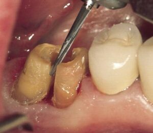
Hemisection
Hemisection is a dental procedure in which one half of a tooth is surgically removed, typically due to significant damage or infection in one root or portion of the tooth. It is usually performed on multi-rooted teeth, such as molars, and aims to preserve the remaining healthy portion of the tooth while eliminating the diseased or compromised part.
Here's a detailed explanation of the hemisection procedure:
- Indications:
- Hemisection is indicated when one root or portion of a multi-rooted tooth is extensively damaged or infected, making it non-restorable through conventional means like fillings or root canal therapy.
- Common reasons for hemisection include advanced periodontal disease, root fractures, extensive decay, or trauma to one root of a multi-rooted tooth.
- Preparation:
- Before the procedure, the dentist will conduct a thorough examination of the affected tooth, including clinical assessment and radiographic evaluation, to determine the extent of damage and plan the treatment.
- Local anesthesia is administered to ensure the patient's comfort during the procedure.
- Procedure:
- Tooth isolation: The tooth and surrounding area are isolated using a dental dam or other protective barrier to maintain a clean and sterile environment.
- Tooth sectioning: Using precise dental instruments, the dentist carefully divides the tooth along the furcation (the area where the roots meet) to separate the healthy portion from the diseased or damaged portion.
- Root removal: The affected root or portion of the tooth is surgically removed, along with any associated infection, debris, or damaged tissue. The remaining healthy root(s) and surrounding bone are preserved.
- Root canal therapy: If the remaining root(s) require root canal treatment, it may be performed to remove any remaining infected or inflamed tissue and seal the root canal space.
- Restoration: After root removal and root canal therapy (if needed), the remaining portion of the tooth is restored with a filling material or crown to restore function and aesthetics.
- Postoperative Care:
- Following the procedure, the patient may experience some discomfort or swelling, which can be managed with pain medication and cold compresses applied to the outside of the cheek.
- Patients are advised to follow postoperative instructions provided by the dentist, including dietary restrictions, oral hygiene practices, and any prescribed medications.
- Regular follow-up appointments are scheduled to monitor healing, assess the stability of the remaining tooth structure, and address any concerns or complications.
- Prognosis:
- The success of hemisection depends on various factors, including the extent of damage or infection, the patient's oral hygiene, and compliance with postoperative care instructions.
- With proper care and maintenance, a tooth that has undergone hemisection can continue to function effectively for many years, providing chewing function and aesthetics similar to a natural tooth.
In summary, hemisection is a dental procedure performed to remove one half of a tooth, typically due to significant damage or infection in one root or portion of the tooth. It aims to preserve the remaining healthy portion of the tooth while eliminating the diseased or compromised part, allowing for continued function and stability. If you have a tooth that may require hemisection or if you have any questions about the procedure, it's important to consult with your dentist for a thorough evaluation and personalized treatment plan.