Emergency Dentist NYC
Contact
Hours
- Monday: 9:00am – 9:00pm
- Tuesday: 9:00am – 6:00pm
- Wednesday: 9:00am – 9:00pm
- Thursday: 9:00am – 9:00pm
- Friday: 9:00am – 5:00pm

Emergency Dentist NYC, located in the vibrant city of New York, New York, is your trusted provider of immediate dental care when dental emergencies occur. Committed to delivering prompt relief and comprehensive treatment for urgent dental issues, our practice offers a wide range of emergency services in a welcoming and comfortable environment. Led by a team of experienced emergency dentists, Emergency Dentist NYC strives to restore your oral health and alleviate your discomfort quickly and efficiently.
Urgent Dental Services
Emergency Examinations
- Rapid Assessments: Immediate evaluations to diagnose and address dental emergencies promptly.
- Pain Management: Quick relief from dental pain through effective anesthesia and pain management techniques.
Emergency Dental Treatments
- Emergency Extractions: Prompt removal of severely damaged or infected teeth causing acute pain or discomfort.
- Emergency Root Canal Therapy: Expedited root canal treatment to alleviate pain and save infected teeth.
Immediate Care
Same-Day Appointments
- Flexible Scheduling: Accommodating same-day appointments for patients in need of urgent dental care.
- Walk-In Services: Convenient walk-in availability for immediate assessment and treatment of dental emergencies.
Efficient Treatment
- Rapid Response: Prompt attention from skilled emergency dentists to address urgent dental needs without delay.
- Streamlined Procedures: Expedited processes to minimize waiting times and efficiently manage dental emergencies.
Patient Care
Compassionate Approach
- Empathetic Staff: Compassionate and understanding dental professionals dedicated to providing comfort and support during stressful situations.
- Clear Communication: Transparent communication about treatment options, costs, and expectations to empower patients to make informed decisions about their dental care.
Aftercare Support
- Post-Treatment Guidance: Detailed instructions and guidance on post-procedure care and pain management to promote optimal healing and recovery.
- Follow-Up Care: Scheduled follow-up appointments to monitor progress, address any concerns, and ensure the successful resolution of dental emergencies.

Veneers
Dental veneers are thin, custom-made shells crafted from tooth-colored materials, such as porcelain or composite resin, that are bonded to the front surface of teeth to improve their appearance and enhance smile aesthetics. Veneers are a popular cosmetic dental treatment option for correcting a variety of dental imperfections and achieving a brighter, more uniform smile. Here's an overview of dental veneers and their key aspects:
- Purpose of Dental Veneers:
- Dental veneers are primarily used to address cosmetic concerns and improve the overall appearance of the smile. They can effectively conceal or correct various dental imperfections, including:
- Discolored or stained teeth that do not respond to teeth whitening treatments.
- Teeth that are chipped, cracked, or worn down.
- Misaligned, uneven, or irregularly shaped teeth.
- Gaps or spaces between teeth.
- Minor crowding or overlapping of teeth.
- Veneers can provide a natural-looking and durable solution for enhancing the size, shape, color, and symmetry of teeth, resulting in a more attractive and confident smile.
- Types of Dental Veneers:
- Porcelain Veneers: Porcelain veneers are custom-crafted shells made from high-quality dental porcelain that closely resembles the appearance of natural tooth enamel. Porcelain veneers are highly durable, stain-resistant, and can produce lifelike results with exceptional translucency and light reflection.
- Composite Resin Veneers: Composite resin veneers are fabricated from tooth-colored composite material that is directly bonded to the teeth and sculpted to achieve the desired shape and appearance. While composite veneers are more affordable and can be completed in a single dental visit, they may be less durable and prone to staining compared to porcelain veneers.
- Treatment Process:
- The dental veneer procedure typically involves multiple steps, starting with a comprehensive dental examination and consultation to assess the patient's oral health, discuss treatment goals, and determine the suitability of veneers.
- During the preparation phase, a small amount of enamel may be removed from the front surface of the teeth to create space for the veneers and ensure a proper fit. In some cases, minimal or no tooth preparation may be required for no-prep or minimal-prep veneers.
- Impressions or digital scans of the teeth are then taken and sent to a dental laboratory, where skilled technicians fabricate custom veneers tailored to the patient's unique dental anatomy, specifications, and desired aesthetic outcome.
- Once the veneers are ready, they are carefully bonded to the front surface of the teeth using a strong dental adhesive or resin cement. The dentist will make any necessary adjustments to ensure proper fit, alignment, and bite before finalizing the bonding process.
- After the veneers are bonded in place, they are polished to achieve a smooth surface and natural appearance, and any excess adhesive is removed. Patients are provided with postoperative instructions and guidelines for maintaining their new veneers and optimizing long-term results.
- Benefits of Dental Veneers:
- Veneers offer numerous benefits for individuals seeking to enhance their smile and address cosmetic dental concerns, including:
- Improved aesthetics and smile confidence.
- Natural-looking results that blend seamlessly with surrounding teeth.
- Long-lasting durability and resistance to staining and discoloration (especially porcelain veneers).
- Minimal tooth preparation and conservative treatment approach.
- Customizable design and shade selection to achieve desired smile goals.
- Immediate transformation of smile imperfections with minimal downtime or recovery.
- Care and Maintenance:
- With proper care and maintenance, dental veneers can provide years of reliable performance and aesthetic appeal. Patients are advised to:
- Practice good oral hygiene habits, including regular brushing, flossing, and routine dental check-ups.
- Avoid biting or chewing on hard objects, such as ice, pens, or fingernails, to prevent damage to the veneers.
- Limit consumption of staining foods and beverages, such as coffee, tea, red wine, and tobacco products, to preserve the appearance of the veneers.
- Wear a protective mouthguard during sports or activities that pose a risk of dental injury.
- Follow any additional instructions or recommendations provided by their dentist for maintaining optimal oral health and veneer longevity.
In summary, dental veneers offer a versatile and effective solution for transforming smiles and achieving a more youthful, attractive appearance. By addressing a wide range of cosmetic dental concerns with natural-looking and durable restorations, veneers empower individuals to smile with confidence and enjoy the benefits of a beautiful and radiant smile.
Cementoblastoma
A cementoblastoma is a rare benign odontogenic tumor that arises from the cementoblasts, which are cells responsible for forming cementum, a specialized calcified tissue that covers the roots of teeth. Cementoblastomas typically occur in association with the roots of teeth, particularly the mandibular (lower) molars, and are characterized by the formation of a well-defined radiopaque (dense) mass attached to the root surface.
Here are some key points about cementoblastomas:
- Etiology: The exact cause of cementoblastomas is not fully understood, but they are thought to arise from aberrant proliferation or differentiation of cementoblasts within the periodontal ligament, the connective tissue that anchors teeth to the surrounding bone. Cementoblastomas are considered true neoplasms (tumors) rather than developmental anomalies, as they consist of proliferating cellular elements rather than disorganized tissue.
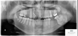
- Clinical Presentation: Cementoblastomas typically present as a painless, slow-growing swelling or mass associated with the roots of the affected tooth. The lesion is usually discovered incidentally on dental radiographs taken for unrelated reasons. On radiographic imaging, cementoblastomas appear as a well-circumscribed radiopaque mass attached to the root surface, often with a characteristic "halo" or radiolucent rim surrounding the lesion.
- Symptoms: In most cases, cementoblastomas are asymptomatic and do not cause pain or discomfort. However, if the lesion grows large enough, it may cause displacement or resorption of adjacent teeth, leading to localized swelling, tooth mobility, or changes in occlusion (bite). Rarely, cementoblastomas may be associated with symptoms such as toothache, facial swelling, or paresthesia (numbness or tingling) if they impinge on adjacent nerves or tissues.
- Diagnosis: Diagnosis of cementoblastomas is typically based on clinical and radiographic findings. Dental X-rays, including periapical, panoramic, or occlusal views, can help visualize the size, shape, and location of the lesion within the jaw. In some cases, additional imaging studies such as CT scans or MRI may be necessary to further evaluate the extent of the lesion and its relationship to surrounding structures.
- Treatment: Treatment of cementoblastomas usually involves surgical removal of the lesion, along with the affected tooth and surrounding periodontal tissues. The procedure is typically performed by an oral and maxillofacial surgeon and may involve extraction of the affected tooth and enucleation (surgical removal) of the tumor while preserving the surrounding bone and adjacent teeth. Following surgical removal, the prognosis for cementoblastomas is excellent, with low rates of recurrence reported.
In summary, cementoblastomas are rare benign odontogenic tumors that arise from the cementoblasts and are typically associated with the roots of teeth. While usually asymptomatic, cementoblastomas may require surgical intervention for removal if they cause symptoms or complications. Early detection and appropriate management are important for achieving favorable outcomes and preserving oral health.