Invisalign Center of NYC
The Invisalign Center of NYC, located in the bustling city of New York, is a premier destination for state-of-the-art orthodontic treatment. Specializing in Invisalign clear aligner therapy, the center offers a modern and convenient approach to achieving straighter teeth and a confident smile. Patients at the Invisalign Center of NYC receive personalized care from a team of skilled orthodontic professionals dedicated to delivering outstanding results.
Orthodontic Services
Invisalign Treatment
- Customized Treatment Plans: Each patient receives a personalized treatment plan tailored to their unique orthodontic needs and smile goals.
- Advanced Technology: Utilizing cutting-edge digital scanning technology to create precise 3D models of the teeth for custom Invisalign aligners.
- Transparent Aligners: Invisalign clear aligners are virtually invisible, providing a discreet and comfortable orthodontic solution.
Comprehensive Care
- Initial Consultation: Thorough examination and discussion of treatment options to determine if Invisalign is the right choice for the patient.
- Progress Monitoring: Regular check-ups to monitor progress and ensure the treatment is progressing as planned.
- Refinements: Fine-tuning the treatment plan as needed to achieve the desired results.
Patient Experience
Convenience and Comfort
- Flexible Appointments: Accommodating scheduling options to fit the busy lifestyles of patients in the heart of NYC.
- Virtual Consultations: Convenient online consultations for initial assessments and treatment planning.
- Comfortable Treatment: Invisalign aligners are smooth and comfortable to wear, with no sharp edges or wires to cause irritation.
Educational Resources
- Smile Simulation: Utilizing advanced software to provide patients with a preview of their smile transformation before starting treatment.
- Oral Hygiene Guidance: Education on proper oral hygiene practices to maintain healthy teeth and gums during treatment.
- Supportive Team: A team of knowledgeable and caring orthodontic professionals available to answer questions and provide guidance throughout the treatment process.
Dentinogenesis Imperfecta
Dentinogenesis imperfecta (DI) is a hereditary genetic disorder that affects the development and formation of dentin, one of the primary tissues that make up teeth. It is characterized by abnormal dentin structure and composition, leading to weakened and discolored teeth that are prone to fracture, wear, and sensitivity. Dentinogenesis imperfecta is typically inherited as an autosomal dominant trait, meaning that a child has a 50% chance of inheriting the condition if one of their parents carries the mutated gene.
Here are some key points about dentinogenesis imperfecta:
- Types: Dentinogenesis imperfecta is classified into three main types based on clinical and genetic features:
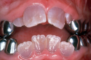
- Type I: Also known as classic or hereditary opalescent dentinogenesis imperfecta, this type is the most common and severe form of the condition. It is characterized by translucent or opalescent (bluish-gray) teeth with bulbous crowns, narrow roots, and severe attrition (wear) of the enamel. Type I DI is caused by mutations in the DSPP (dentin sialophosphoprotein) gene, which encodes a protein involved in dentin formation.
- Type II: Also known as coronal dentinogenesis imperfecta, this type is characterized by similar dental abnormalities as type I DI but with less severe enamel involvement. Teeth may appear yellow-brown or amber in color and may be more resistant to fracture compared to type I DI. Type II DI is also caused by mutations in the DSPP gene.
- Type III: Also known as Brandywine type dentinogenesis imperfecta, this type is characterized by similar dental abnormalities as type II DI but with additional skeletal abnormalities such as short stature and joint laxity. Type III DI is caused by mutations in the DSPP gene as well.
- Clinical Presentation: Dentinogenesis imperfecta typically presents with a distinctive appearance of the teeth, including opalescent or discolored enamel, bulbous crowns, and attrition of the enamel exposing the underlying dentin. The teeth may appear translucent or amber in color, and the enamel may chip or fracture easily due to its weakened structure. Individuals with dentinogenesis imperfecta may also experience dental sensitivity, pulp exposure, and increased risk of dental caries and infections.
- Diagnosis: Diagnosis of dentinogenesis imperfecta is based on clinical and radiographic findings, including characteristic dental abnormalities such as opalescent or discolored enamel, bulbous crowns, and narrowed pulp chambers. Dental X-rays may reveal thin and bulbous roots, obliteration of the pulp chambers, and dentin defects such as taurodontism (enlarged pulp chambers) or pulpal calcifications. Genetic testing may be performed to confirm the diagnosis and identify the underlying genetic mutation responsible for the condition.
- Treatment: Treatment of dentinogenesis imperfecta focuses on preserving tooth structure, preventing complications, and improving oral function and aesthetics. Management options may include dental restorations such as crowns, veneers, or composite fillings to protect and reinforce weakened teeth, extraction of severely affected teeth followed by prosthetic replacement, endodontic therapy (root canal treatment) for teeth with pulp exposure or infection, and preventive measures such as fluoride therapy and meticulous oral hygiene to reduce the risk of dental caries and infections.
In summary, dentinogenesis imperfecta is a hereditary genetic disorder characterized by abnormal development and structure of dentin, resulting in weakened and discolored teeth that are prone to fracture, wear, and sensitivity. Early diagnosis and appropriate dental management are essential for preserving tooth structure, preventing complications, and improving oral function and aesthetics in individuals with dentinogenesis imperfecta.
Tooth Discoloration
Tooth discoloration is a common dental concern characterized by changes in the color of the teeth, which can occur due to various factors affecting the enamel, dentin, or pulp tissues. Discoloration can manifest as stains, spots, or overall changes in tooth color, ranging from yellow or brown discoloration to gray, black, or blue hues. Understanding the causes and types of tooth discoloration can help guide appropriate management and treatment options.
Here are some key points about tooth discoloration:
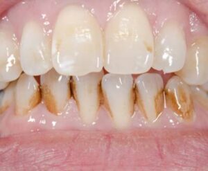
- Extrinsic Discoloration: Extrinsic discoloration occurs when stains or pigments accumulate on the surface of the enamel, typically due to external factors such as dietary habits, oral hygiene practices, or environmental exposures. Common causes of extrinsic discoloration include:
- Food and beverages: Consumption of deeply pigmented foods and drinks such as coffee, tea, red wine, berries, or curry can lead to extrinsic staining of the teeth over time.
- Tobacco use: Smoking or chewing tobacco products can result in extrinsic staining and discoloration of the teeth, often manifesting as brown or yellowish stains on the enamel surface.
- Poor oral hygiene: Inadequate brushing, flossing, or professional dental cleanings can allow plaque and tartar buildup on the teeth, contributing to extrinsic discoloration and surface stains.
- Intrinsic Discoloration: Intrinsic discoloration occurs when changes in the internal structure or composition of the tooth tissues affect the overall color of the tooth from within. Common causes of intrinsic discoloration include:
- Developmental factors: Genetic anomalies, enamel defects, or excessive fluoride intake during tooth development can lead to intrinsic discoloration of the teeth.
- Aging: As individuals age, the enamel layer may wear down, exposing the underlying dentin, which is naturally yellowish or brown in color, resulting in intrinsic discoloration and a darker appearance of the teeth.
- Trauma or injury: Dental trauma, such as falls, accidents, or sports injuries, can disrupt blood flow to the developing teeth or cause internal hemorrhage, leading to intrinsic discoloration or tooth darkening over time.
- Medications: Certain medications or medical treatments, such as tetracycline antibiotics (when taken during tooth development), chemotherapy, or radiation therapy, can cause intrinsic discoloration of the teeth, particularly in children or adolescents.
- Combined Discoloration: In some cases, tooth discoloration may result from a combination of extrinsic and intrinsic factors, where surface stains accumulate on the enamel surface, while underlying structural changes contribute to internal discoloration.
- Diagnosis: Diagnosis of tooth discoloration involves a thorough clinical examination, assessment of medical and dental history, and identification of potential causative factors. Dental X-rays and other diagnostic tests may be performed to evaluate the internal structure of the teeth and rule out underlying pathology or anomalies contributing to discoloration.
- Treatment: Treatment of tooth discoloration depends on the underlying causes and severity of the discoloration. Management options may include:
- Professional dental cleanings to remove surface stains and plaque buildup.
- Teeth whitening treatments to lighten and brighten the enamel surface.
- Dental restorations such as veneers or crowns to cover or mask intrinsic discoloration.
- Prevention measures such as proper oral hygiene practices, dietary modifications, and lifestyle changes to minimize the risk of tooth discoloration.
In summary, tooth discoloration can result from a variety of factors affecting the enamel, dentin, or pulp tissues. Diagnosis and treatment of tooth discoloration require a comprehensive approach to address both surface stains and underlying structural changes, with management options ranging from professional dental cleanings and teeth whitening to dental restorations and preventive measures to maintain oral health and aesthetics.


