Kids Only Dental
Contact
Hours
- Monday: 1:00am – 9:00pm
- Tuesday: 9:00am – 6:00pm
- Wednesday: 1:00am – 9:00pm
- Thursday: 9:00am – 9:00pm
- Friday: 9:00am – 5:00pm

Kids Only Dental, located in New York, NY, is a premier pediatric dental practice dedicated exclusively to the dental needs of children. The clinic is known for its child-friendly environment, state-of-the-art facilities, and a team of highly trained animal dental professionals who provide compassionate and specialized care to ensure a positive dental experience for every child.
Dental Services
Preventive Dentistry
- Comprehensive Oral Exams: Thorough examinations to monitor and maintain oral health, with a focus on early detection of dental issues.
- Routine Cleanings: Regular cleanings to prevent cavities and promote healthy teeth and gums.
- Fluoride Treatments: Strengthening treatments to protect children’s teeth from decay.
- Dental Sealants: Protective coatings applied to the chewing surfaces of molars to prevent cavities.
Specialized Pediatric Dental Care
- Restorative Dentistry: Fillings and crowns to repair cavities and restore damaged teeth in children.
- Orthodontic Assessments: Early evaluations and referrals for orthodontic treatment to correct dental alignment and bite issues.
- Emergency Dental Care: Prompt and effective treatment for dental emergencies, such as toothaches, broken teeth, or dental trauma.
- Behavior Management: Techniques to help children feel comfortable and at ease during dental visits, including sedation dentistry options for anxious patients.
Educational Programs
- Oral Hygiene Education: Teaching children proper brushing and flossing techniques to encourage good oral hygiene habits.
- Nutrition Counseling: Guidance on healthy eating habits that support dental health.
- Parental Support: Resources and advice for parents to help them care for their children’s dental needs at home.
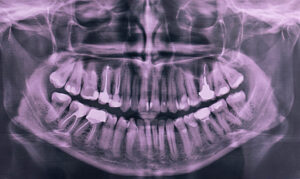
X-Rays
Dental X-rays, also known as dental radiographs, are diagnostic imaging techniques used by dentists to visualize and assess the internal structures of the teeth, jaws, and surrounding tissues that are not visible during a regular dental examination. Here's an overview of dental X-rays and their key aspects:
- Purpose of Dental X-Rays:
- Dental X-rays serve various purposes in dental care, including:
- Detecting tooth decay (cavities) between teeth or under existing fillings.
- Evaluating the health of the tooth roots and surrounding bone.
- Assessing the development and eruption of permanent teeth in children and adolescents.
- Detecting abnormalities, such as cysts, tumors, or impacted teeth.
- Planning and monitoring orthodontic treatment (braces or aligners).
- Evaluating the extent of dental trauma or injury.
- Assessing the bone density and structure for dental implant placement.
- Types of Dental X-Rays:
- There are several types of dental X-rays commonly used in dental practice, each serving a specific purpose:
- Bitewing X-rays: Used to detect cavities between the back teeth (molars and premolars) and assess the fit of dental fillings.
- Periapical X-rays: Provide detailed images of the entire tooth, including the crown, root, and surrounding bone.
- Panoramic X-rays: Capture a broad view of the entire mouth, including the jaws, teeth, sinuses, and temporomandibular joints (TMJ).
- Occlusal X-rays: Focus on a specific area of the mouth to evaluate the development of teeth or detect abnormalities.
- Cephalometric X-rays: Used in orthodontics to assess the relationship between the teeth, jaws, and facial structures.
- Radiation Safety and Dose:
- Dental X-rays emit very low levels of radiation, and modern X-ray equipment and techniques minimize radiation exposure to patients.
- Dentists adhere to strict radiation safety protocols, such as using lead aprons and thyroid collars to shield the patient's body from unnecessary exposure.
- The benefits of dental X-rays in diagnosing and preventing oral health problems far outweigh the minimal risks associated with radiation exposure.
- Procedure and Technique:
- During a dental X-ray procedure, the patient is positioned in a chair or standing next to the X-ray machine, and protective aprons or shields are placed to cover areas not being imaged.
- The X-ray machine is positioned close to the area of interest, and the dentist or radiology technician instructs the patient to hold still and bite down on a film or digital sensor placed inside the mouth.
- The X-ray machine emits a small burst of radiation, which penetrates the tissues and creates an image of the teeth and surrounding structures on the film or sensor.
- Digital X-ray technology allows for instant image capture and viewing on a computer monitor, reducing the time and effort required to develop traditional X-ray films.
- Interpretation and Diagnosis:
- After acquiring dental X-ray images, the dentist carefully examines and interprets the radiographic findings to assess the patient's oral health status and formulate an appropriate treatment plan.
- Dental X-rays help dentists identify dental issues early, allowing for timely intervention and treatment to prevent further complications.
- Dentists may compare current X-ray images with previous ones to monitor changes in the patient's oral health over time and evaluate the effectiveness of treatment interventions.
- Patient Education and Communication:
- Dentists often use dental X-ray images as visual aids to educate patients about their oral health condition, treatment options, and preventive measures.
- Patient communication and informed consent are essential aspects of dental X-ray procedures, and dentists discuss the benefits, risks, and necessity of X-rays with their patients before obtaining consent for imaging.
In summary, dental X-rays are valuable diagnostic tools that enable dentists to visualize and assess the internal structures of the teeth and jaws, aiding in the diagnosis, treatment planning, and monitoring of various oral health conditions. By utilizing appropriate X-ray techniques and adhering to radiation safety protocols, dentists ensure the safe and effective use of X-rays in dental practice.
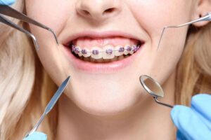
Orthodontic Treatment
Orthodontic treatment is a specialized dental procedure designed to correct misaligned teeth and jaws. It focuses on improving the alignment of teeth and bite to enhance oral function, aesthetics, and overall dental health. Here's a breakdown of orthodontic treatment:
- Purpose:
- Orthodontic treatment aims to address various dental issues, including:
- Crooked or crowded teeth.
- Gaps or spaces between teeth.
- Overbite, underbite, crossbite, or open bite.
- Misaligned jaws or improper dental arch alignment.
- By correcting these issues, orthodontic treatment not only enhances the appearance of the smile but also improves chewing function, speech clarity, and overall oral health.
- Orthodontic Appliances:
- Braces: Traditional metal braces consist of brackets attached to the teeth connected by wires and bands. They apply gentle pressure to gradually shift teeth into the desired position.
- Clear Aligners: These are transparent, removable trays made of smooth plastic that are custom-fit to the patient's teeth. Brands like Invisalign® use clear aligners to discreetly straighten teeth.
- Functional Appliances: These devices, such as headgear or palate expanders, are used to modify jaw growth patterns and correct bite discrepancies in growing children.
- Retainers: After completing orthodontic treatment, retainers are often prescribed to maintain the new tooth positions and prevent relapse.
- Treatment Process:
- Orthodontic treatment typically begins with a comprehensive examination, including X-rays, photographs, and impressions of the teeth.
- The orthodontist develops a personalized treatment plan based on the patient's specific needs and goals.
- Regular appointments are scheduled to adjust braces or monitor progress with clear aligners. The duration of treatment varies depending on the complexity of the case and the chosen orthodontic method.
- Patients are instructed on proper oral hygiene practices and may receive dietary advice to maintain healthy teeth and gums during treatment.
- Benefits:
- Improved Aesthetics: Orthodontic treatment enhances the appearance of the smile by aligning teeth and correcting bite issues, leading to increased self-confidence and a more attractive smile.
- Enhanced Function: Properly aligned teeth improve bite function, making it easier to chew food and speak clearly. This can also alleviate issues like jaw pain or discomfort.
- Reduced Risk of Dental Problems: Straight teeth are easier to clean, reducing the risk of cavities, gum disease, and other dental issues associated with crowded or misaligned teeth.
- Overall Oral Health: Orthodontic treatment contributes to better long-term oral health by creating a balanced bite and improving the stability of the teeth and supporting structures.
- Post-Treatment Care:
- After orthodontic treatment, patients may need to wear retainers to maintain the results and prevent teeth from shifting back.
- Regular dental check-ups and cleanings are essential for monitoring oral health and ensuring that the teeth remain stable and healthy.
In summary, orthodontic treatment offers numerous benefits beyond just cosmetic improvement. By correcting misalignments and bite issues, it helps patients achieve optimal oral health, function, and aesthetics, leading to a lifetime of confident smiles and improved overall well-being.