Manhattan Maxillofacial Surgery Group
Contact
Hours
- Monday: 9:00am – 9:00pm
- Tuesday: 9:00am – 6:00pm
- Wednesday: 9:00am – 9:00pm
- Thursday: 9:00am – 9:00pm
- Friday: 9:00am – 5:00pm

Manhattan Maxillofacial Surgery Group, located in the heart of New York City, is a leading practice specializing in oral and maxillofacial surgery. The group is renowned for its state-of-the-art facilities, cutting-edge technology, and a team of highly skilled animal surgeons dedicated to providing exceptional care. Patients receive personalized, compassionate treatment tailored to their specific surgical needs.
Surgical Services
Oral Surgery
- Wisdom Teeth Extraction: Safe and effective removal of impacted or problematic wisdom teeth.
- Dental Implants: Expert placement of dental implants to replace missing teeth and restore oral functionality.
- Tooth Extractions: Removal of damaged or decayed teeth, with options for sedation to ensure patient comfort.
Maxillofacial Surgery
- Jaw Surgery (Orthognathic Surgery): Corrective surgery to address jaw alignment issues, improve function, and enhance facial aesthetics.
- Treatment of Facial Trauma: Comprehensive care for facial injuries, including fractures and lacerations.
- Reconstructive Surgery: Rebuilding facial structures affected by trauma, disease, or congenital conditions.
Advanced Procedures
- TMJ Disorders: Diagnosis and surgical treatment of temporomandibular joint disorders to relieve pain and improve jaw function.
- Bone Grafting: Advanced bone grafting techniques to prepare the jaw for dental implants or to repair bone loss.
- Oral Pathology: Diagnosis and surgical treatment of diseases affecting the mouth, jaws, and related structures.
Patient Care
Consultation and Planning
- Comprehensive Evaluations: Detailed assessments to create personalized treatment plans tailored to each patient’s needs.
- Advanced Imaging: Use of cutting-edge imaging technology, including 3D scans, for precise diagnosis and treatment planning.
- Pre- and Post-Operative Care: Thorough guidance and support before and after surgery to ensure optimal recovery and outcomes.
Comfort and Support
- Patient Education: Detailed explanations of procedures and recovery processes to help patients feel informed and confident.
- Comfort Management: Options for sedation and anesthesia to ensure patient comfort and reduce anxiety during procedures.
- Follow-Up Care: Regular follow-up appointments to monitor healing and address any concerns promptly.
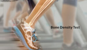
Bone Density Evaluation
Bone density evaluation, also known as bone density testing or bone densitometry, is a medical procedure used to measure the strength and density of bones, typically in the hip, spine, or forearm. This assessment helps healthcare providers diagnose osteoporosis, assess fracture risk, and monitor the effectiveness of treatment for bone-related conditions. Here's an overview of bone density evaluation:
- Indications:
- Bone density evaluation is recommended for individuals at risk of osteoporosis or fractures, including postmenopausal women, older adults, individuals with a family history of osteoporosis, and those with certain medical conditions or taking medications that affect bone health.
- It may also be recommended for individuals who have experienced a fracture or have other risk factors for osteoporosis, such as low body weight, smoking, excessive alcohol consumption, or a sedentary lifestyle.
- Bone Densitometry Techniques:
- Dual-energy X-ray absorptiometry (DXA or DEXA): This is the most commonly used technique for measuring bone density. It involves using low-dose X-rays to scan the hip, spine, or forearm and assess bone mineral density (BMD). DXA scans are quick, non-invasive, and provide precise measurements of bone density.
- Quantitative ultrasound (QUS): This technique measures bone density using sound waves instead of X-rays. It is often used as a screening tool and may be performed at the heel or wrist.
- Peripheral dual-energy X-ray absorptiometry (pDXA): This portable device measures bone density at peripheral sites such as the wrist, heel, or finger.
- Preparation:
- Preparation for a bone density evaluation is typically minimal. Patients may be advised to wear loose, comfortable clothing and avoid wearing metal objects or jewelry that may interfere with the scan.
- In some cases, patients may be instructed to refrain from taking calcium supplements or certain medications prior to the test, as they can affect the results.
- Procedure:
- During a DXA scan, the patient lies on a table while a scanner arm passes over the body, emitting low-dose X-rays. The procedure is painless and usually takes about 10 to 30 minutes to complete.
- For QUS or pDXA, the patient may be asked to place the body part being scanned (such as the heel or wrist) against a small device that emits sound waves or X-rays.
- Interpretation of Results:
- The results of the bone density evaluation are typically reported as a T-score, which compares the patient's bone density to that of a healthy young adult of the same gender. A T-score of -1 or above is considered normal, while scores between -1 and -2.5 indicate low bone density (osteopenia), and scores below -2.5 indicate osteoporosis.
- The healthcare provider will interpret the results of the bone density evaluation in conjunction with other clinical factors, such as age, gender, medical history, and fracture risk, to assess the patient's overall bone health and determine appropriate management strategies.
- Follow-up and Monitoring:
- Depending on the results of the bone density evaluation, the healthcare provider may recommend lifestyle modifications, dietary changes, weight-bearing exercises, and/or medications to help maintain or improve bone health.
- Follow-up bone density evaluations may be recommended periodically to monitor changes in bone density over time and assess the effectiveness of treatment interventions.
Bone density evaluation is a valuable tool for assessing bone health, diagnosing osteoporosis, and guiding treatment decisions to reduce fracture risk and improve overall bone health. If you are at risk of osteoporosis or fractures, or if you have concerns about your bone health, it's important to discuss with your healthcare provider whether a bone density evaluation is appropriate for you.
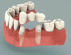
Bridges
Dental bridges are fixed dental prostheses used to replace one or more missing teeth by bridging the gap between natural teeth or dental implants. They consist of one or more artificial teeth, known as pontics, which are attached to adjacent natural teeth or implants for support. Bridges are custom-made to match the color, size, and shape of the patient's natural teeth, restoring both function and aesthetics to the smile.
Here are the key aspects of dental bridges:
- Types of Bridges:
- Traditional Bridges: These are the most common type of bridges and consist of one or more pontics attached to dental crowns, which are cemented onto the natural teeth adjacent to the gap.
- Cantilever Bridges: In cases where there is only one adjacent tooth next to the gap, a cantilever bridge may be used. This type of bridge is anchored to only one natural tooth or implant.
- Maryland Bridges (Resin-Bonded Bridges): Maryland bridges use metal or porcelain wings bonded to the backs of adjacent natural teeth, eliminating the need for dental crowns. They are often used for replacing front teeth.
- Procedure:
- The process of getting a dental bridge typically involves two or more dental appointments.
- During the initial appointment, the dentist prepares the abutment teeth, which are the natural teeth adjacent to the gap, by removing a portion of their enamel to accommodate the dental crowns.
- Impressions of the prepared teeth and the surrounding dental structures are taken to fabricate the custom-made bridge in a dental laboratory.
- A temporary bridge may be placed to protect the prepared teeth while the final bridge is being fabricated.
- Once the bridge is ready, the temporary bridge is removed, and the final bridge is cemented into place, restoring function and aesthetics to the smile.
- Benefits of Dental Bridges:
- Restoration of Function: Dental bridges restore the ability to chew and speak properly, which may be compromised by missing teeth.
- Aesthetic Improvement: Bridges fill in the gaps left by missing teeth, improving the appearance of the smile and restoring facial symmetry.
- Prevention of Tooth Movement: By filling in the space left by missing teeth, bridges help prevent neighboring teeth from shifting out of position.
- Longevity: With proper care and maintenance, dental bridges can last for many years, providing a durable and cost-effective solution for tooth replacement.
- Post-Treatment Care:
- Patients with dental bridges should practice good oral hygiene habits, including brushing twice a day, flossing daily, and attending regular dental check-ups and cleanings.
- Avoiding hard or sticky foods and habits like biting on pens or fingernails can help prolong the life of the bridge and prevent damage.
- Regular monitoring by a dentist is essential to ensure the bridge remains stable and functional over time.
In summary, dental bridges offer a reliable and effective solution for replacing missing teeth, restoring both function and aesthetics to the smile. With proper care and maintenance, bridges can provide patients with a long-lasting and natural-looking tooth replacement option.