Stony Brook Medicine
Contact
Hours
- Monday: 9:00am – 9:00pm
- Tuesday: 9:00am – 6:00pm
- Wednesday: 9:00am – 9:00pm
- Thursday: 9:00am – 9:00pm
- Friday: 9:00am – 5:00pm

Stony Brook Medicine, located in Stony Brook, New York, is a renowned healthcare institution providing a wide array of medical and dental services. Known for its state-of-the-art facilities and cutting-edge technology, Stony Brook Medicine is staffed by a dedicated team of highly trained animal medical professionals committed to delivering compassionate and innovative care to all patients.
Medical Services
General Medicine and Surgery
- Emergency Services: Available 24/7, featuring the latest emergency medical technology and highly skilled animal medical personnel.
- Inpatient and Outpatient Care: Comprehensive services including internal medicine, cardiology, neurology, orthopedics, and more.
- Robotic Surgery: Advanced minimally invasive procedures using cutting-edge robotic technology for precision and faster recovery times.
Specialized Departments
- Rheumatology: Expert care for arthritis, autoimmune diseases, and musculoskeletal disorders.
- Dermatology: Comprehensive treatment for skin conditions, including eczema, psoriasis, and skin cancer.
- Pulmonology: Advanced care for respiratory conditions, including asthma, COPD, and lung infections.
- Geriatrics: Specialized care for elderly patients, focusing on maintaining health, independence, and quality of life.
Dental Services
General Dentistry
- Preventive Care: Routine checkups, cleanings, and education to maintain oral health.
- Sealants and Fluoride Treatments: Protective treatments to prevent decay, especially in children.
Specialized Dental Care
- Oral and Maxillofacial Pathology: Diagnosis and treatment of diseases affecting the oral and maxillofacial regions.
- Implant Dentistry: Expert placement of dental implants to replace missing teeth and restore functionality.
- Periodontics: Advanced treatment for gum diseases and other conditions affecting the tissues surrounding the teeth.
- Orthodontics: Comprehensive orthodontic treatments for children and adults to correct dental alignment and bite issues, including braces and clear aligners.
- Sleep Dentistry: Specialized care for dental issues related to sleep disorders, such as sleep apnea.
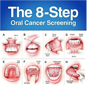
Oral Cancer Screening
Oral cancer screening is a preventive healthcare measure aimed at detecting signs and symptoms of oral cancer or precancerous lesions in the mouth, lips, tongue, gums, throat, or adjacent tissues at an early stage when treatment is most effective. Here's an overview of oral cancer screening and its key aspects:
- Importance of Oral Cancer Screening:
- Oral cancer is a serious and potentially life-threatening condition that can affect anyone, regardless of age, gender, or lifestyle factors. Early detection of oral cancer greatly improves the chances of successful treatment and survival.
- Oral cancer screening allows dentists and healthcare providers to identify suspicious lesions or abnormalities in the oral cavity that may indicate the presence of oral cancer or precancerous changes.
- Regular oral cancer screening is particularly important for individuals at higher risk, including tobacco users (smokers and smokeless tobacco users), heavy alcohol consumers, individuals with a family history of oral cancer, and those with human papillomavirus (HPV) infection.
- Screening Methods and Techniques:
- Oral cancer screening may be performed as part of a routine dental examination or as a standalone procedure during a visit to the dentist or healthcare provider.
- The screening process typically involves a visual examination of the oral cavity, lips, tongue, gums, throat, and adjacent tissues to look for any abnormalities, such as lumps, bumps, ulcers, red or white patches, or other suspicious lesions.
- In some cases, additional diagnostic tests or imaging studies, such as toluidine blue staining, brush biopsy, tissue biopsy, or imaging modalities like X-rays, CT scans, or MRI scans, may be recommended to further evaluate suspicious lesions and confirm or rule out the presence of cancer.
- Clinical Signs and Symptoms of Oral Cancer:
- During the oral cancer screening, the dentist or healthcare provider will look for the following signs and symptoms that may raise suspicion of oral cancer:
- Persistent mouth ulcers or sores that do not heal within two weeks.
- Red or white patches (leukoplakia or erythroplakia) on the oral mucosa.
- Unexplained swelling, lumps, or thickening of tissues in the mouth or neck.
- Pain, numbness, or difficulty swallowing (dysphagia).
- Chronic hoarseness or changes in voice quality.
- Persistent sore throat, ear pain, or jaw stiffness.
- Unexplained bleeding or numbness in the mouth.
- Risk Factors for Oral Cancer:
- Several factors increase the risk of developing oral cancer, including:
- Tobacco Use: Smoking cigarettes, cigars, pipes, or using smokeless tobacco products (chewing tobacco, snuff) greatly increases the risk of oral cancer.
- Heavy Alcohol Consumption: Excessive alcohol consumption, especially when combined with tobacco use, significantly raises the risk of oral cancer.
- HPV Infection: Certain strains of human papillomavirus (HPV), particularly HPV-16 and HPV-18, have been linked to an increased risk of oral and oropharyngeal cancers.
- Sun Exposure: Prolonged exposure to ultraviolet (UV) radiation from the sun or tanning beds increases the risk of lip cancer.
- Age and Gender: Oral cancer tends to occur more frequently in older adults over the age of 50 and is more common in men than women.
- Preventive Measures and Follow-up:
- While oral cancer screening can help detect early signs of oral cancer, prevention is key to reducing the risk of developing the disease.
- Individuals can reduce their risk of oral cancer by avoiding tobacco use, moderating alcohol consumption, practicing good oral hygiene, eating a healthy diet rich in fruits and vegetables, and minimizing exposure to known risk factors.
- Regular dental check-ups and oral cancer screenings are essential for early detection and prompt treatment of oral cancer. Patients should follow up with their dentist or healthcare provider if they notice any changes or abnormalities in their oral health between screenings.
In summary, oral cancer screening is a vital component of preventive healthcare aimed at detecting oral cancer and precancerous lesions at an early stage when treatment is most effective. By undergoing regular screenings, individuals can reduce their risk of developing oral cancer and improve their chances of successful treatment and recovery.
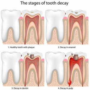
Tooth Decay
Tooth decay, also known as dental caries or cavities, is a common dental problem characterized by the breakdown of tooth structure due to the demineralization of enamel and dentin by acids produced by bacteria in the mouth. It is one of the most prevalent chronic diseases worldwide and can lead to pain, infection, and tooth loss if left untreated. Tooth decay occurs when the natural balance of bacteria in the mouth is disrupted, leading to the formation of plaque, a sticky film of bacteria and food particles that adheres to the teeth. The bacteria in plaque metabolize sugars and carbohydrates from food, producing acids that attack the tooth enamel and eventually lead to the formation of cavities.
Here are some key points about tooth decay:
- Causes:
- Bacteria: Oral bacteria, particularly Streptococcus mutans and Lactobacillus species, play a crucial role in the development of tooth decay by metabolizing sugars and carbohydrates from food and producing acids that demineralize tooth enamel.
- Diet: Consumption of sugary or carbohydrate-rich foods and beverages, such as candy, soda, fruit juice, and refined carbohydrates, provides fuel for bacteria in the mouth and contributes to the formation of plaque and tooth decay.
- Poor oral hygiene: Inadequate brushing, flossing, and tongue cleaning allow plaque to accumulate on the teeth, increasing the risk of tooth decay.
- Dry mouth: Reduced saliva flow, often caused by medications, medical conditions, or mouth breathing, can impair the natural cleansing and remineralization of teeth, increasing susceptibility to tooth decay.
- Genetics: Genetic factors may influence an individual's susceptibility to tooth decay, including the composition of saliva, tooth enamel structure, and immune response to bacteria.
- Stages:
- Initial demineralization: In the early stages of tooth decay, acids produced by bacteria in plaque attack the tooth enamel, causing demineralization and weakening of the enamel surface.
- Formation of cavities: As the enamel continues to demineralize, it may eventually break down, leading to the formation of small holes or cavities in the tooth surface.
- Progression of decay: If left untreated, tooth decay can progress deeper into the tooth, reaching the dentin layer and eventually the dental pulp, leading to pain, infection, and possible tooth loss.
- Symptoms:
- Tooth sensitivity: Sensitivity to hot, cold, sweet, or acidic foods and beverages is often an early sign of tooth decay.
- Toothache: Persistent or intermittent tooth pain, particularly when chewing or biting down, may indicate advanced tooth decay or infection.
- Visible holes or pits in the teeth: Cavities may be visible as dark spots, holes, or pits on the surface of the teeth.
- Discoloration: Discoloration or darkening of the teeth may occur as decay progresses and the enamel becomes thinner.
- Diagnosis:
- Diagnosis of tooth decay is typically based on a dental examination, evaluation of symptoms, and diagnostic tests such as dental X-rays or visual inspection with dental instruments.
- Your dentist will examine the teeth for signs of decay, including visible cavities, discoloration, or softening of the enamel.
- Dental X-rays may be taken to assess the extent of decay and detect cavities between the teeth or beneath the enamel surface.
- Treatment:
- Treatment of tooth decay aims to remove the decayed tissue, restore the tooth structure, and prevent further damage. Treatment options may include:
- Dental fillings: In cases of mild to moderate decay, your dentist may remove the decayed tissue and fill the cavity with a dental filling material such as composite resin, amalgam, or glass ionomer cement.
- Dental crowns: For more extensive decay or weakened teeth, your dentist may recommend placing a dental crown to restore the strength, function, and appearance of the tooth.
- Root canal therapy: If decay reaches the dental pulp and causes infection or inflammation, root canal therapy may be necessary to remove the infected tissue, clean and disinfect the root canal space, and seal it to prevent further infection.
- Tooth extraction: In cases of severe decay or irreparable damage, tooth extraction may be necessary to remove the affected tooth and prevent spread of infection to surrounding tissues.
- Prevention:
- To prevent tooth decay, it's important to:
- Practice good oral hygiene habits, including regular brushing with fluoride toothpaste, flossing, and tongue cleaning.
- Limit consumption of sugary or carbohydrate-rich foods and beverages that contribute to plaque formation and tooth decay.
- Drink water or chew sugar-free gum after meals to help rinse away food particles and neutralize acids in the mouth.
- Visit your dentist regularly for professional cleanings and check-ups to detect and treat tooth decay early.
In summary, tooth decay is a common dental problem characterized by the breakdown of tooth structure due to acid erosion by bacteria