1-888-3289

Odontogenic Keratocyst

At Barnet Health, we offer diagnosis and treatment of odontogenic keratocyst.

Odontogenic Keratocyst

At Barnet Health, we offer diagnosis and treatment of odontogenic keratocyst.

What is it?

An odontogenic keratocyst (OKC), also known as keratocystic odontogenic tumor (KCOT), is a benign but locally aggressive cystic lesion that originates from the dental lamina or remnants of the dental lamina. It is one of the most common types of cysts that affect the jaws and is often associated with developmental anomalies such as nevoid basal cell carcinoma syndrome (Gorlin-Goltz syndrome).

Here are some key points about odontogenic keratocysts:

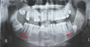
  1. Origin: Odontogenic keratocysts arise from the remnants of the dental lamina, which is an embryonic structure involved in tooth development. They most commonly occur in the posterior mandible (lower jaw), particularly around the angle and ramus areas, but can also affect the maxilla (upper jaw) and other regions of the jaws. These cysts typically present as well-defined radiolucent lesions on dental radiographs.
  2. Clinical Presentation: Odontogenic keratocysts are often asymptomatic and may be discovered incidentally on routine dental radiographs. However, they can grow to large sizes and cause swelling, expansion of the jawbone, displacement of adjacent teeth, and paresthesia (numbness) of the lower lip or chin if they impinge on the inferior alveolar nerve. In some cases, odontogenic keratocysts may cause pain or discomfort if they become infected or associated with other pathologic conditions.
  3. Histopathology: Microscopically, odontogenic keratocysts are characterized by a thin, stratified epithelial lining with a distinct basal layer and a parakeratinized or orthokeratinized surface. The epithelial lining exhibits a high mitotic index and a tendency for proliferation and invagination into the surrounding connective tissue, leading to the formation of daughter cysts or satellite cysts. The presence of a characteristic palisading or corrugated basal cell layer is a hallmark feature of odontogenic keratocysts.
  4. Treatment: The management of odontogenic keratocysts typically involves surgical intervention to remove the cystic lesion and prevent recurrence. This may include enucleation (complete removal of the cystic lining) with or without curettage (scraping) of the surrounding bone, marsupialization (creation of a surgical window to decompress the cyst), or resection of the affected jaw segment in cases of extensive or aggressive lesions. Adjunctive measures such as chemical cauterization with Carnoy’s solution or cryotherapy may be employed to reduce the risk of recurrence by destroying residual epithelial remnants.
  5. Recurrence: Odontogenic keratocysts have a high recurrence rate compared to other types of jaw cysts, with reported recurrence rates ranging from 10% to 62% following surgical treatment. Recurrence is thought to be attributed to the presence of satellite cysts, daughter cysts, or residual epithelial rests that are not completely removed during surgery. Close long-term follow-up with clinical and radiographic examinations is essential for monitoring postoperative healing and detecting early signs of recurrence.

In summary, odontogenic keratocyst is a benign but locally aggressive cystic lesion that originates from the remnants of the dental lamina. It commonly affects the jaws and may present with swelling, expansion, or displacement of adjacent structures. Surgical removal is the mainstay of treatment, but recurrence rates are relatively high, necessitating long-term follow-up and surveillance.

Free E-Book!

Learn the Causes and Solutions to Gum Disease!